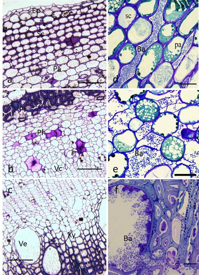
Fig 4. Light micrographs of healthy and infected kiwifruit bark.
(a–c), Transverse sections of healthy bark, showing typical anatomical tissues consisting of a typical epidermis (Ep), sclerenchyma (Sc), parenchyma (Pa), phloem fiber (Pf), phloem (Ph), vascular cambium (Vc), xylem (Xy), and vessel (Ve). Bar = 100 μm. (d–f), transverse sections of inoculated bark. (d), bacterial cells (Ba) in intercellular spaces of parenchyma; (e), bacterial cells in phloem cells and intercellular spaces; (f), bacterial cells tightly packed within xylem vessels. Bar = 20 μm.

