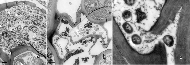
Fig 5. Transmission electron micrographs of cross sections of kiwifruit twigs infected with the GFPuv-labeled Psa 5 DAI.
(a), Bacterial cells in intercellular spaces of the parenchyma. Bar = 2 μm; (b), bacterial colonization in parenchyma cells where the cell walls are ruptured and organelles have disintegrated. Bar = 2 μm; (c), bacterial colonization in intercellular spaces where the middle lamella tended to be ruptured. Bar = 0.5 μm.

