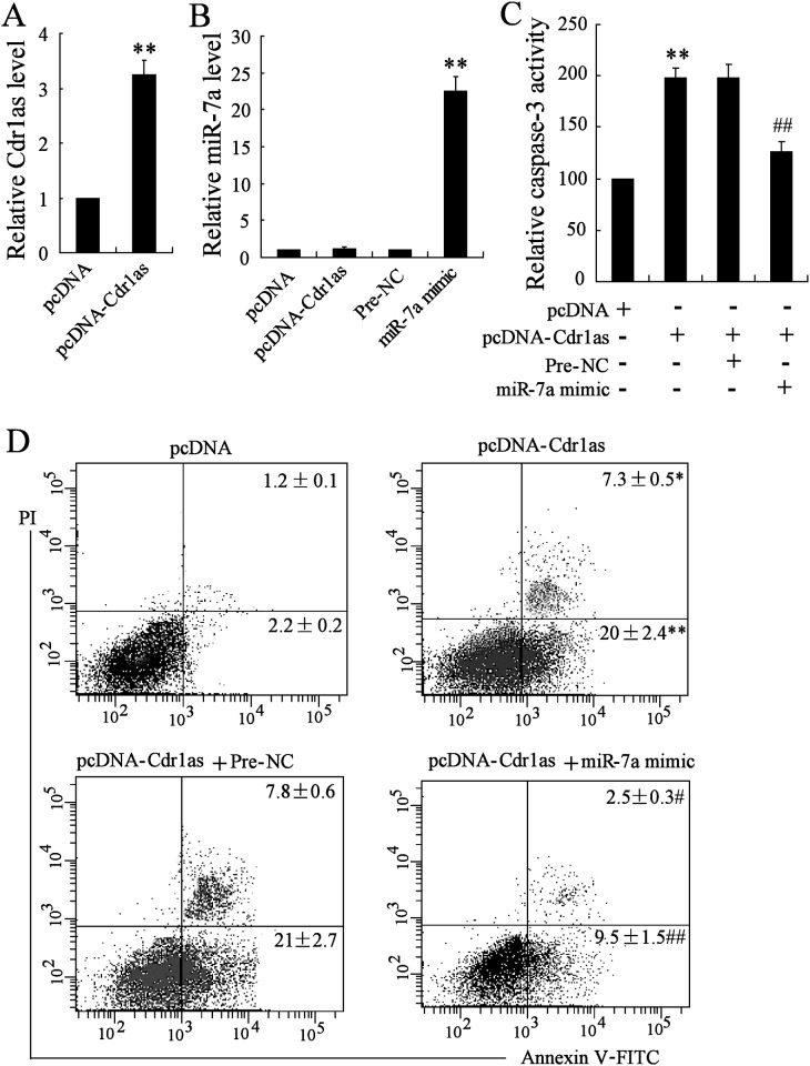
Fig 3. MiR-7a overexpression reversed Cdr1as-induced apoptosis of MCM cells.
MCM cells were transfected with pcDNA-Cdr1as or miR-7a mimic for the overexpression of Cdr1as and miR-7a respectively, with pcDNA or Pre-NC as the respective negative control. After the transfection, Cdr1as or miR-7a expression level was confirmed by qRT-PCR. Analysis for caspase-3 activity and cell apoptosis were performed by the colorimetric method and Annexin V/PI staining respectively. A. Expression of Cdr1as. B. Expression of miR-7a. C. Relative caspase-3 activity under different conditions. D. Representative flow cytometry of apoptosis of MCM cells. The percentage of apoptotic cells with positive Annexin V signal were analyzed and compared accordingly. n = 3, *P < 0.05, **P<0.01: compared to cells transfected with pcDNA (A, C, D) or Pre-NC (B) as control; #P < 0.05, ##P < 0.01: compared to cells co-transfected with pcDNA-Cdr1as and Pre-NC.

