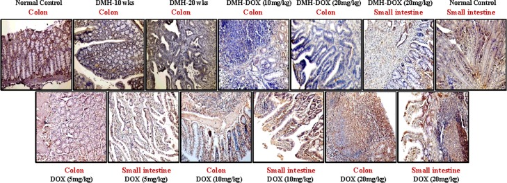
Fig 6. Immunohistochemistry for colonic and small intestinal tissues from normal control, DMH-alone, DMH-DOX and DOX-alone-treated rats.
Immunostaining with anti-p53. The signal for protein is represented by brown color due to DAB and blue signal due to hematoxylin counterstain. Magnification ×200; (n = 24).

