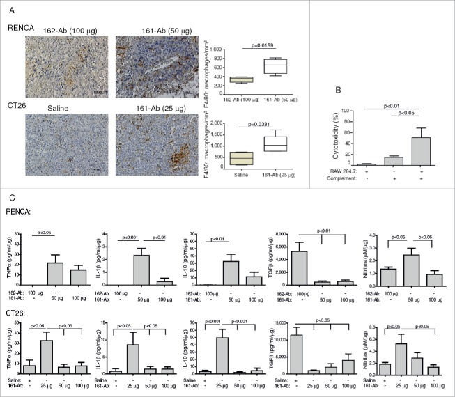
Figure 6.
161-Ab increases macrophage infiltration, mediates tumor cell cytotoxicity, and changes tumor microenvironment. (A) Representative images of immunohistochemical staining for F4/80 in RENCA and CT26 tumor sections. Bar size for RENCA and CT26 tumors is 50 μm. Number of macrophages per mm2 was quantified (n = 4–5). (B) CT26 cells (5×10⁴ cells) were labeled with Cell Tracker, incubated in vitro with 1.28 ng/mL of the 161-Ab for 6 h, in the presence of complement (diluted 1:50), RAW 264.7 cells at a ratio of 2:1, or both. Percent cytotoxicity were determined as described in the methods (n = 4). (C) Cytokine concentration in tumor lysates were determined by ELISA, and normalized to the amount of total protein in each sample (n = 4–5).

