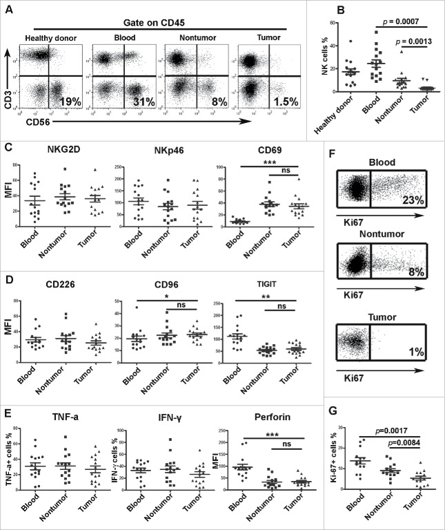
Figure 2.
NK cell percentage, phenotype, functions and proliferation in GC patients. For the measurement of intracellular cytokine production, mononuclear cells were isolated from blood, nontumoral tissues and intratumoral tissues. Per 2 × 10⁶ mononuclear cells were cultured in 500 uL of complete medium with 2 uL Leukocyte Activation Cocktail (BD Bioscience) in 48 well plates in a 37°C humidified CO2 incubator for 4 h. Following activation, cells were stained with surface markers, fixed and permeabilized with IntraPre Reagent (Beckman Coulter), and finally stained with anti-TNF-α, anti-IFN-γ, anti-perforin and anti-granzyme B. (A, B) Representative image and statistical data of the percentages of total NK cells in lymphocytes as determined by flow cytometry. (C, D) Statistical results of the surface receptors expressed on the NK cells. (E) Statistical results of the assay for IFNγ, TNF-α and granzyme B secreted by the NK cells. (F, G) Representative image and statistical data of the percentage of Ki-67+ cells among the total NK cells. Results are expressed as the means ± SEM; * p < 0.05; ** p < 0.01; *** p < 0.001.

