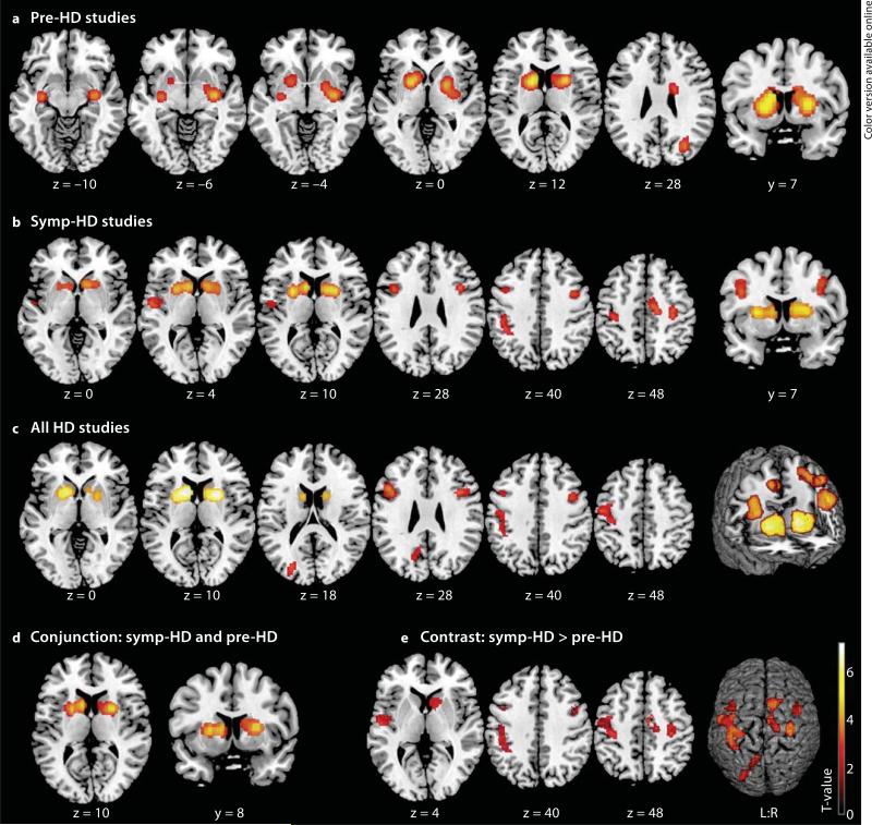
Fig. 1.
Significant clusters from ALE meta-analyses displayed on a template brain (coordinates in Montreal Neurological Institute space, left is left). a Regions of volume loss in pre-HD subjects compared to controls were located in the bilateral striatum extending to the amygdala, hippocampus, right pallidum, posterior insula, occipital cortex and left thalamus. b Volume loss in symp-HD studies was observed in bilateral basal ganglia, inferior frontal gyrus and motor cortices (PMC, M1), extending to the left somatosensory cortex (SI, SII), parietal areas (IPC, IPS), insula and right midcingulate cortex. c Atrophy across all HD studies was detected in the same areas, except for the right motor cortex and left SII, and was additionally observed in left occipitoparietal areas. d Conjunction analysis. Common atrophy in pre-HD and symp-HD was found in the bilateral basal ganglia. e Contrast analysis. More pronounced atrophy in symp-HD compared to pre-HD was found in the right basal ganglia, bilateral inferior frontal gyrus and motor cortex, left somatosensory, parietal and occipital areas, insula and right midcingulate cortex.

