
Figure 1.
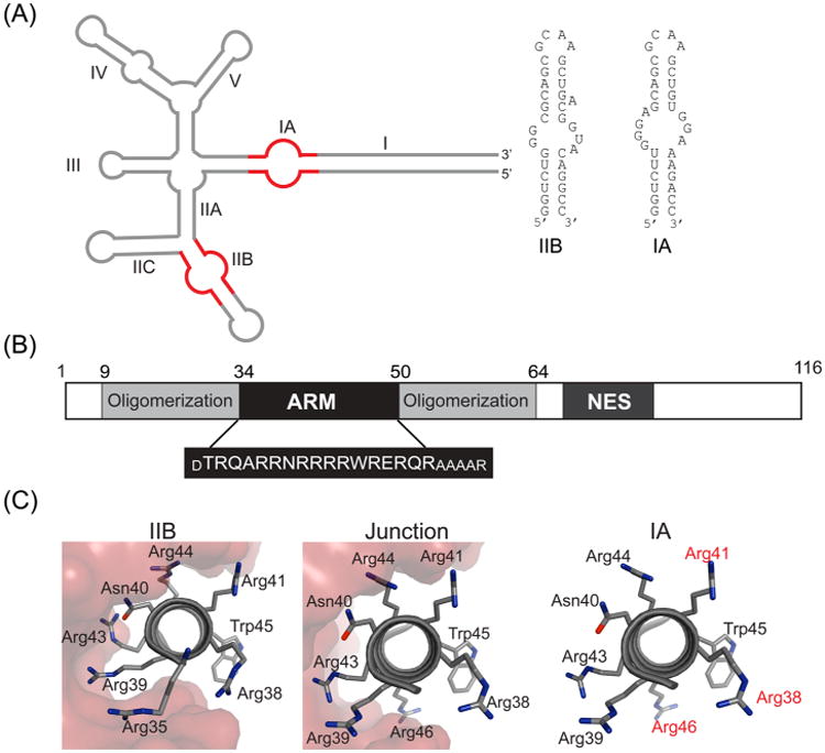
HIV-1 Rev protein and RRE RNA. (A) Secondary structure of HIV-1 RRE15 with stems IIB and IA shown in red and the sequence and secondary structures of the two IIB and IA RNA hairpins, used in this study. Rev assembly nucleates at stem IIB and most likely proceeds via the IIABC junction along stem IIA to stem IA and stem I. (B) Domain organization of HIV-1 Rev with the sequence of the arginine-rich motif (ARM) shown below. The residues in smaller font were added to the ARM sequence to increase its helical content.18,23 (C) Rev–ARM interactions with three known binding sites on the RRE. The same surface of the ARM is used for RNA recognition at IIB and junction sites (shown in red surface representation),10 while a different surface is likely used for IA binding (residues implicated in IA recognition are shown in red).18