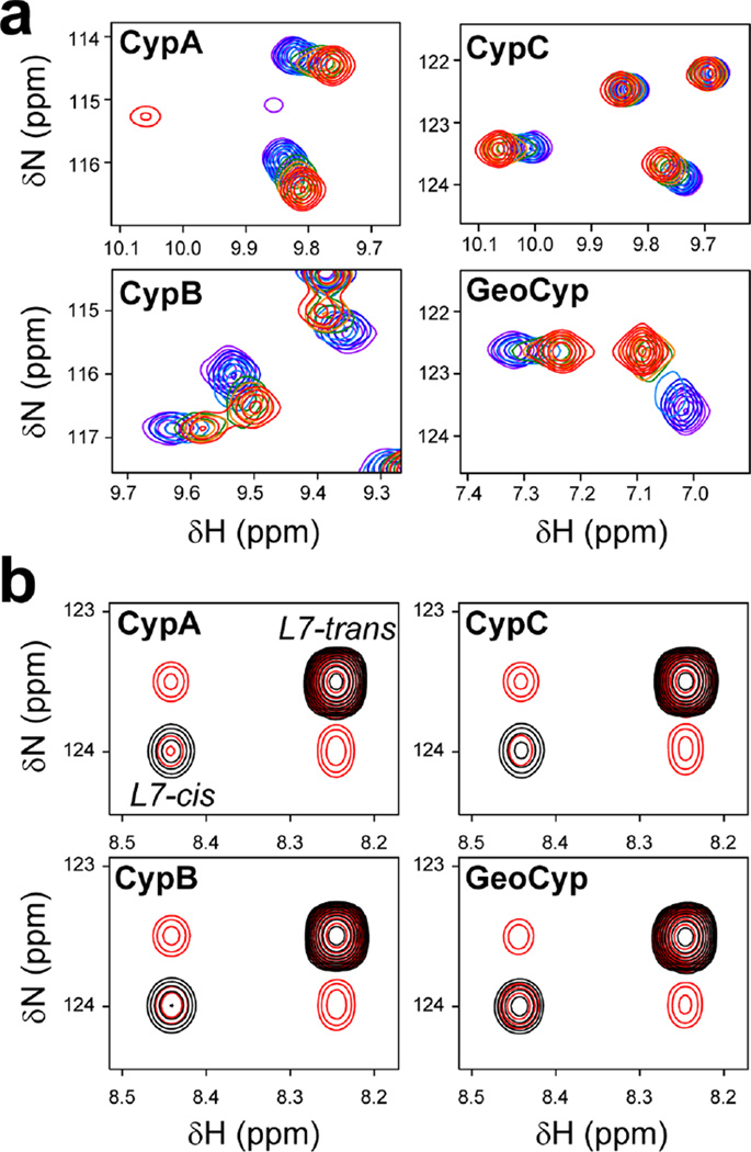
Figure 1.
Functional characterization of cyclophilins. (a) Titrations o the peptide substrate into 15N-labeled cyclophilins. 15N HSQC spectra were collected on 0.5 mM cyclophilin with 0 mM (red), 0.1 mM (orange), 0.2 mM (green), 0.5 mM (cyan), 1 mM (blue), and 2 mM (violet) unlabeled peptide substrate. (b) ZZ-exchange spectra for residue Leu 7, collected with mixing times of 0 ms (black) and 144 ms (red) on 1 mM 15N-labeled peptide with the addition of 20 µM cyclophilin

