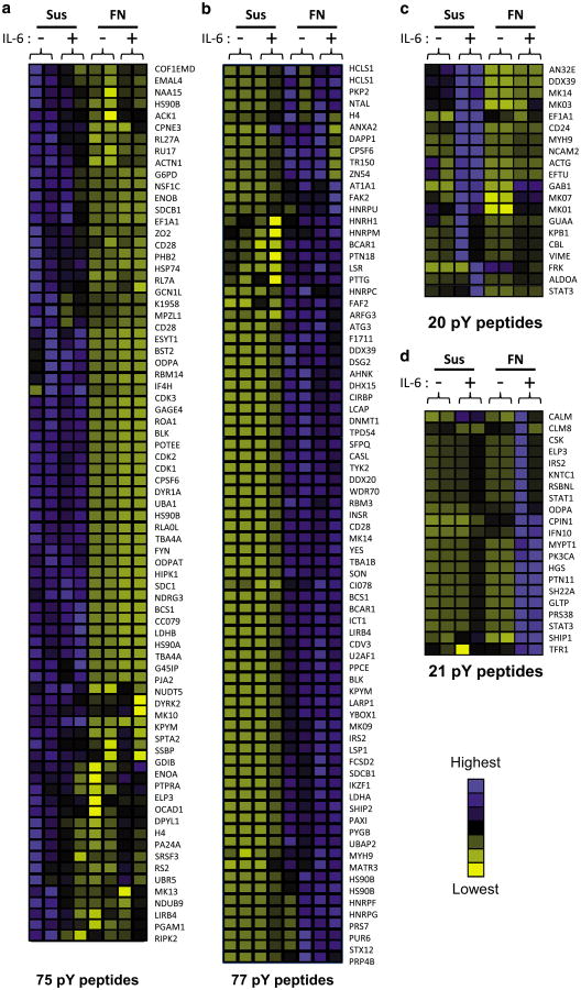
Figure 2.
Adhesion to fibronectin alters the IL-6-induced phosphotyrosine proteome of 8226 myeloma cells. 8226 myeloma cells were grown in suspension (Sus) or adhered to FN plates (FN) for 1 h prior to stimulation with 1 ng/ml IL-6 for 30 min. Lysates from 108 cells per condition were digested with trypsin, and phosphotyrosine peptides were enriched by immunoprecipitation. Peptides are grouped according to changes in relative intensity between sample groups: phosphorylation induced by (a) suspension, (b) adhesion to FN, (c) Sus+IL-6 or (d) adhesion to FN+IL-6. Peptides with increasing relative abundance are indicated by blue shading; decreasing phosphorylation events are indicated by yellow shading. Peptides are identified by gene symbols.

