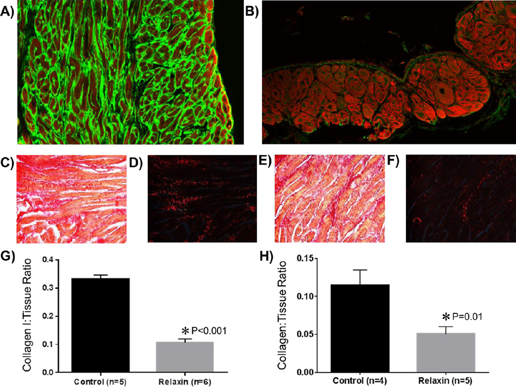
Figure 4. Effect of Relaxin on Atrial Fibrosis.
The top panels are atrial immunohistological sections (20×) of aged rat hearts treated with vehicle (A) or relaxin (B). Phalloidin is represented in red and collagen I in green. The middle panels are aged rat atrial sections (40×) stained with picrosirius red. Dark red staining denotes fibrosis. Images C&D are from vehicle treated aged rats imaged under non-polarized (C) and polarized light (D). Images E&F are from relaxin treated aged rats under non-polarized (E) and polarized light (F). G and H quantify the collagen I or collagen to tissue ratio.

