Figure 2.
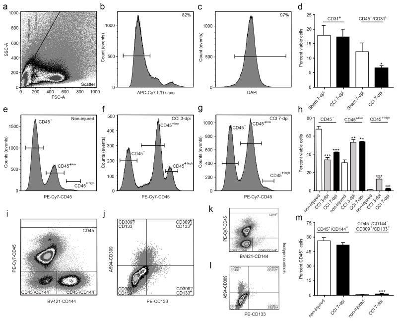
CCI injury leads to differential changes in the percent of CD45− and CD45+ subpopulations of cells in the cortex. (a) Scatter plot shows exclusion of cellular debris. Viable (b) and nucleated (c) cells were selected for using a live/dead stain followed by DAPI staining. (d) No difference was observed in viable CD31+ (PECAM-1) cells between sham and CCI-injured mice at 7 dpi; however, excluding CD45+ cells from the analysis results in a significant decrease in CD45−/CD31+ cvECs. (e–h) CCI injury increased the percent of CD45high (infiltrating leukocytes) and CD45low (residential microglia), while reducing the population of CD45− cells. (i) Scatter plot showing separation of CD144+ (VE-cadherin) cvECs, and CD309+ (VEGFR-2)/CD133+ (Prominin-1) EPCs (j). Scatter plot showing isotype controls for CD45−/CD144+ (k) and CD309+/CD133+ (l) populations. (m) At 7 dpi the percentage of CD45−/CD144+ ECs was not changed whereas the smaller CD309+/CD133+ EPC population was significantly increased. n=3 biological replicates. * p<0.05, ** p<0.01, *** p<0.001 as compared with non-injured mice.

