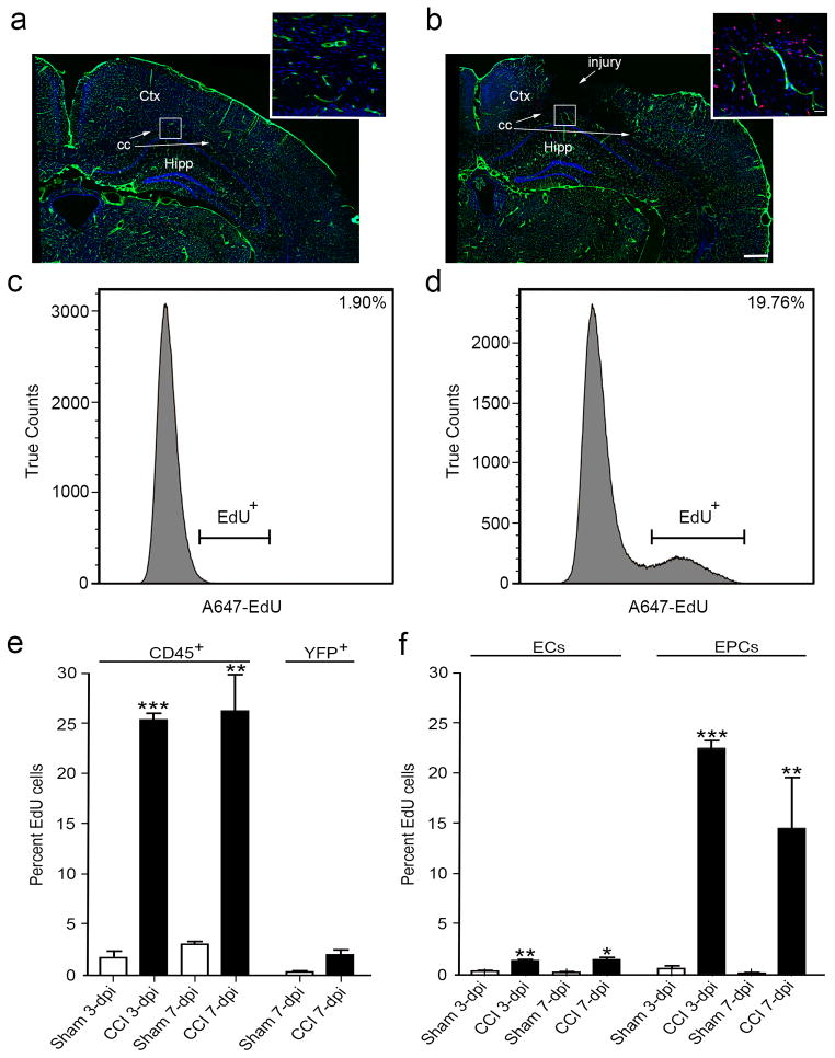
Figure 4.
Proliferating EdU+ infiltrating cells (CD45+), cvECs and EPCs are increased at 3 and 7 dpi. (a) Immunostained coronal brain section of non-injured sham Cgh5-zG (green) cortex shows DAPI staining (blue) and few proliferating cells (red). Inset is a high-magnification image of subcortical layers. (b) Immunostained coronal brain section of CC-injured Cgh5-zG (green) cortex at 3 dpi shows DAPI staining (blue) and greater numbers of proliferating cells (red). Inset is a high-magnification image of subcortical layers in the penumbra. (c, d) Flow cytometry analysis revealed a nearly 20% increase in total viable EdU+ cells at 3 dpi (d) compared with sham (c) mice. (e) CD45+ infiltrating cells were significantly proliferating at 3 and 7 dpi, while YFP+ neurons were not proliferating. (f) cvEC and EPC were significantly proliferating at 3 and 7 dpi. (a, b) n=3; (c–f) n=9–12. ** p<0.01, *** p<0.001 as compared with non-injured sham mice. Ctx, cortex; cc, corpus callosum; Hipp, hippocampus.

