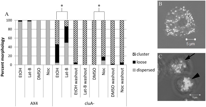
Figure 1.
Analysis of mitochondria distribution in AX4 and cluA− cytoskeleton disrupted cells. (A) AX4 mitochondrial morphology remained dispersed and was unaffected by all treatments. The cluA− clustered mitochondria phenotype was significantly decreased with more loose clusters and dispersed mitochondrial when treated with latrunculin-B (p < 0.0001) or nocodazole (p < 0.0001). (B,C) Examples of mitochondrial distribution. (B) AX4 cell with dispersed mitochondria, (C) cluA− cells where top cell (with arrow) shows loosely clustered mitochondria, bottom cell (with arrowhead) shows a tight cluster. *Indicates significant differences.

