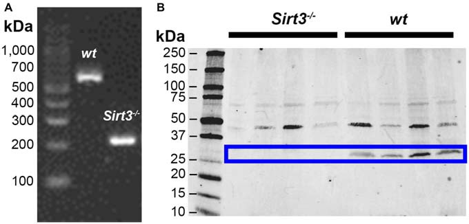
Figure 1.

Representative images showing detection of Sirt3 in the mouse colon by RT-PCR (A) and western blot (B). Tissue from Sirt3−/− mice was used as a negative control. PCR amplification of Sirt3 mRNA (A) shows the expected products at 562 bp for wt Sirt3 and 200 bp for mutant Sirt3 (Sirt3−/−). Western blots for Sirt3 protein (B) show two bands potentially corresponding to the full-length nuclear protein at 44 kDa and the processed mitochondrial form at 28 kDa. However, only the band at 28 kDa is absent in Sirt3−/− mice and this band corresponds to the single known form of mouse Sirt3.
