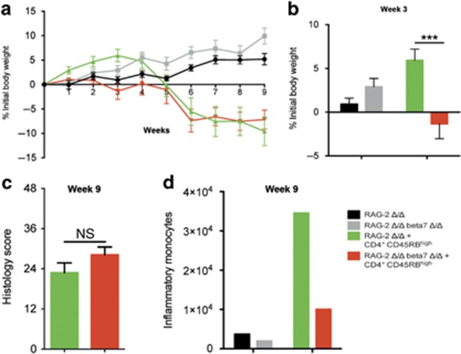
Figure 5.
β7-Integrin on innate immune cells is of minor importance for the chronic stage of T-cell-mediated colitis. Recombination activating gene-2-deficient (RAG-2 Δ/Δ) and RAG-2 Δ/Δ β7-integrin-deficient (RAG-2 Δ/Δ β7Δ/Δ) mice were adoptively transferred with 0.5 × 10⁶ CD4+CD25-CD45RBhi naïve T cells from wild-type mice and assessed for colitis development. Mice were weighed weekly. At 9 weeks after transfer, the mice were killed and analyzed by flow cytometry and histology. Color code: black=RAG-2 Δ/Δ mice that received no cells (control); gray=RAG-2 Δ/Δ β7Δ/Δ mice that received no cells (control); green=RAG-2 Δ/Δ recipient mice that received CD4+CD25−CD45RBhi cells; red=RAG-2 Δ/Δ β7Δ/Δ recipient mice that received CD4+CD25−CD45RBhi cells. (a, b) Body weight as a percent of starting weight of RAG-2 Δ/Δ control mice (black, n=5), RAG-2 Δ/Δ β7Δ/Δ control mice (gray, n=5), RAG-2 Δ/Δ recipient mice (green, n=11), and RAG-2 Δ/Δ β7Δ/Δ recipient mice (red, n=11) over the course of 9 weeks (a) and after 3 weeks (b). (c) Results of histological scoring of sections from RAG-2 Δ/Δ recipient mice (green, n=8) and RAG-2 Δ/Δ β7 integrin Δ/Δ recipient mice (red, n=11) mice after 9 weeks of chronic T-cell-mediated colitis. (d) Representative example of a flow cytometric quantification of inflammatory monocytes per small intestine (SI) after week 9 of chronic T-cell-mediated colitis of RAG-2 Δ/Δ control mice (black), RAG-2 Δ/Δ β7Δ/Δ control mice (gray), RAG-2 Δ/Δ recipient mice (green), and RAG-2 Δ/Δ β7Δ/Δ recipient mice (red). Flow cytometry data are from pooled small intestinal samples (n=5 mice each). The data shown are representative of two independent experiments. Data are presented as mean±s.e.m. Statistical significance was calculated by two-way analysis of variance (ANOVA) with Sidak's post-test and is indicated as follows: ***P<0.001.

