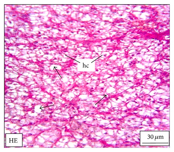
Figure 5.

The histological section of the liver of a M. tengara during the early spawning phase (June) depicting comparatively fewer numbers of fat vacuoles (small black arrows) and slight decrease in the size of the hepatocytes (hc) under experimental condition when the tissues are treated with ZnS nanoparticles at the concentration of 250 μg/L [HE: Haematoxylin-Eosin stain].
