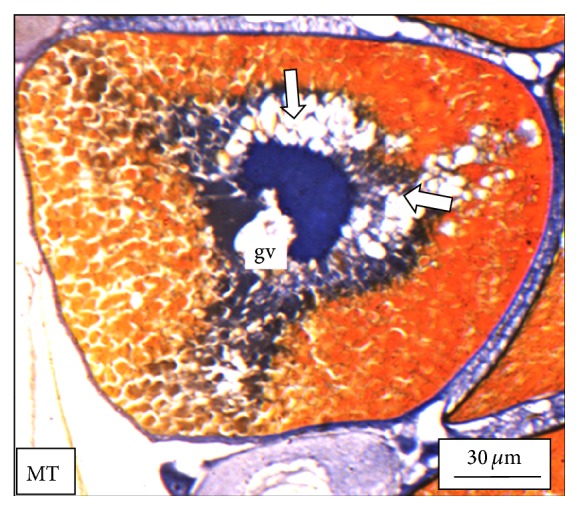
Figure 6.

The histological section of the ovary of a M. tengara during the early spawning phase (June) showing maturing follicles (mf) with impeded yolk deposition. Empty yolk vesicles (solid white arrows) around the basophilic germinal vesicle (gv) under experimental condition when the tissues are treated with ZnS nanoparticles at the concentration of 250 μg/L also support the deposition of the yolk material centripetally [Mallory's triple stain].
