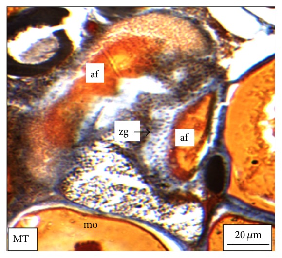
Figure 7.

The histological section of the ovary of a M. tengara during the early spawning phase (June) showing mature oocytes undergoing apoptosis and showing liquefaction of the yolk materials along with hypertrophy of the granulosa cell (zg) [HE: Haematoxylin-Eosin stain].
