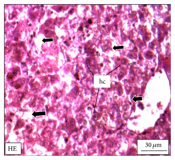
Figure 9.

The histological section of the liver of a M. tengara during the late spawning phase (August) depicting the diffused array of hepatocytes (hc) that is diminished in size but bearing prominent nuclei. The fat vacuoles are totally absent; instead some conspicuous empty spaces (black solid arrows) are generated in between the hepatocytes when the tissues are treated with ZnS nanoparticles at the concentration of 500 μg/L [HE: Haematoxylin-Eosin stain].
