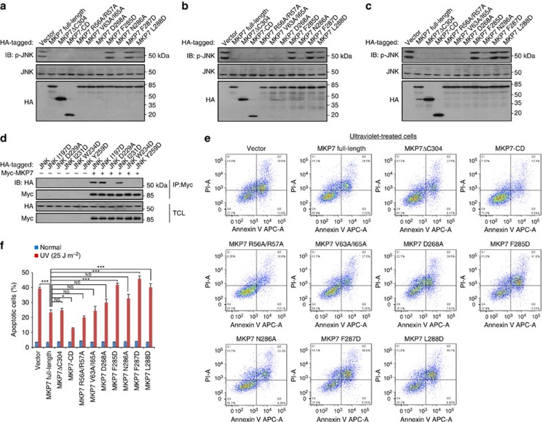
Figure 6. FXF-motif is critical for controlling the phosphorylation of JNK and ultraviolet-induced apoptosis.
(a–c) FXF-motif is essential for the dephosphorylation of JNK by MKP7. HEK293T cells were infected with lentiviruses expressing MKP7 and its mutants (1.0 μg). After 36 h infection, cells were untreated in a, stimulated with 30 μM etoposide for 3 h in b or irradiated with 25 J m−2 ultraviolet light at 30 min before lysis in c. Whole-cell extracts were then immunoblotted with antibody indicated. Shown is a typical immunoblot for phosphorylated JNK from three independent experiments. (d) F-site is required for JNK1 to interact with MKP7. HEK293T cells were co-transfected with MKP7 full-length (1.0 μg) and JNK1 (wild type or mutants as indicated, 1.0 μg). At 16 h post transfection, cells were lysed. Whole-cell extracts were then immunoprecipitated with antibody against Myc for MKP7; immunobloting was carried out with antibodies indicated. IP, immunoprecipitation; TCL, total cell lysate. Shown is a typical result from three independent experiments. (e) Effect of MKP7 (wild type or mutants) expression on ultraviolet-induced apoptosis. HeLa cells were infected with lentiviruses expressing MKP7 full-length and its mutants. At 36 h post infection, cells were irradiated with 25 J m−2 ultraviolet light and collected at 6 h after irradiation. Cells were then subjected to flow cytometry analysis. Apoptotic cells were determined by Annexin-V-APC/PI staining. The results using Annexin-V stain for membrane phosphatidylserine eversion, combined with propidium iodide (PI) uptake to evaluate cells whose membranes had been compromised. Staining with both Annexin-V and PI indicate apoptosis (upper right quadrant). The values shown in the lower left, and upper right quadrants of each panel represent the percentage of viable, and apoptotic cells, respectively. All results are representative of three independent experiments. (f) Statistical analysis of apoptotic cells (mean±s.e.m., n=3), *P<0.05, ***P<0.001 (ANOVA followed by Tukey's test). NS, not significant.

