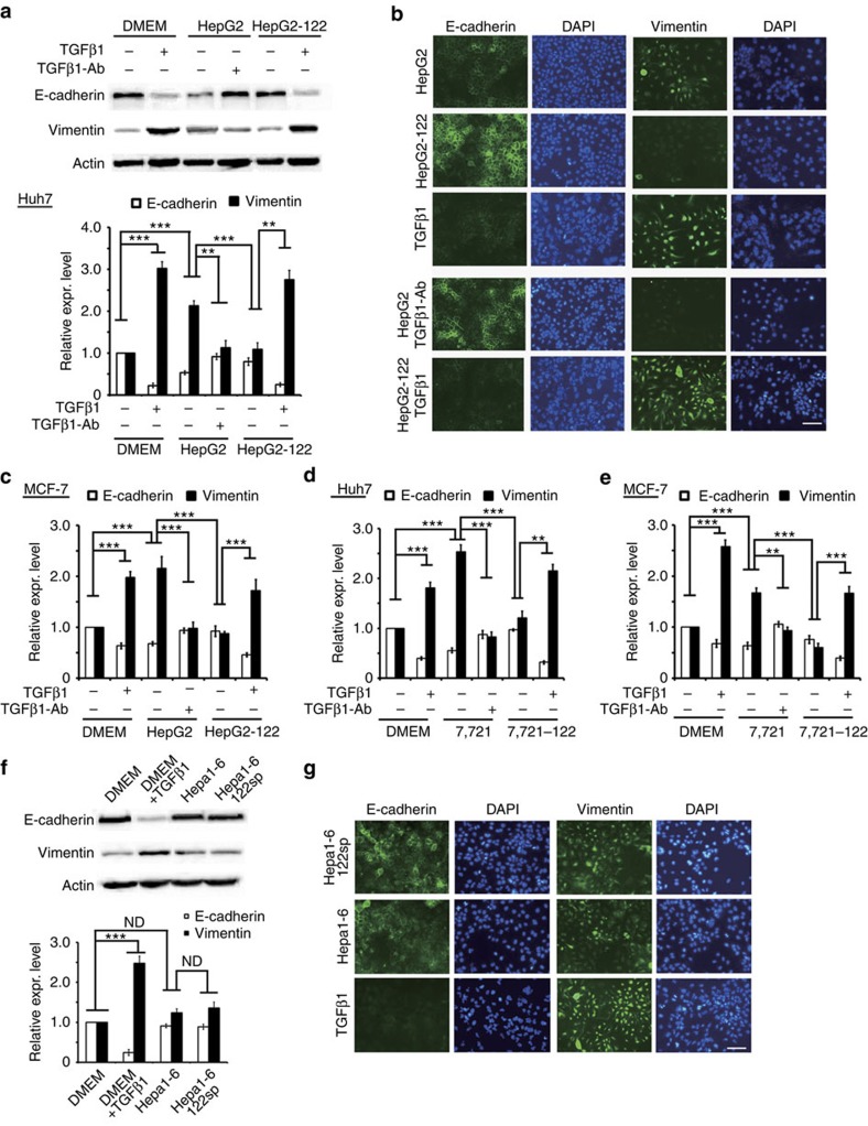
Figure 4. Differential targeting of TGFβ1/TGFβR1 is the underlying reason for the distinct impact of miR-122 on EMT in human or mouse cells.
(a) Western blot analysis of E-cadherin and vimentin levels in Huh7 treated as indicated. HepG2 and HepG2-122 stands for the supernatant of HepG2 and HepG2-122 cells, respectively. Quantitative analysis is shown on the right. Three independent repeats are performed in each experiment. (b) E-cadherin and vimentin were used as markers of EMT in Huh7 cells treated as indicated. 4,6-Diamidino-2-phenylindole (DAPI) staining was used to detect nuclei. Scale bar, 50 μm. (c) Quantitative analysis of E-cadherin and vimentin levels in MCF-7 treated as indicated. HepG2 and HepG2-122 stands for the supernatant of HepG2 and HepG2-122 cells by western blot, respectively. Three independent repeats are performed in each experiment. (d) Quantitative analysis of E-cadherin and vimentin levels in Huh7 treated as indicated. 7721 and 7721-122 stands for the supernatant of SMC-7721 and SMC-7721-122 cells by western blot, respectively. Three independent repeats are performed in each experiment. (e) Quantitative analysis of E-cadherin and vimentin levels in MCF-7 treated as indicated. 7721 and 7721-122 stands for the supernatant of SMC-7721 and SMC-7721-122 cells by western blot, respectively. Three independent repeats are performed in each experiment. (f) Western blot analysis of E-cadherin and vimentin levels in Huh7 treated as indicated. Hepa1-6 and Hepa1-6-122sp stands for the supernatant of Hepa1-6 and Hepa1-6-122sp cells, respectively. Three independent repeats are performed in each experiment. (g) E-cadherin and vimentin were used as markers of EMT in Huh7 cells treated as indicated. DAPI staining was used to detect nuclei. Scale bar, 50 μm. Error bars, ±s.d. **P<0.01; ***P<0.001 by two-sided Student's t-test.

