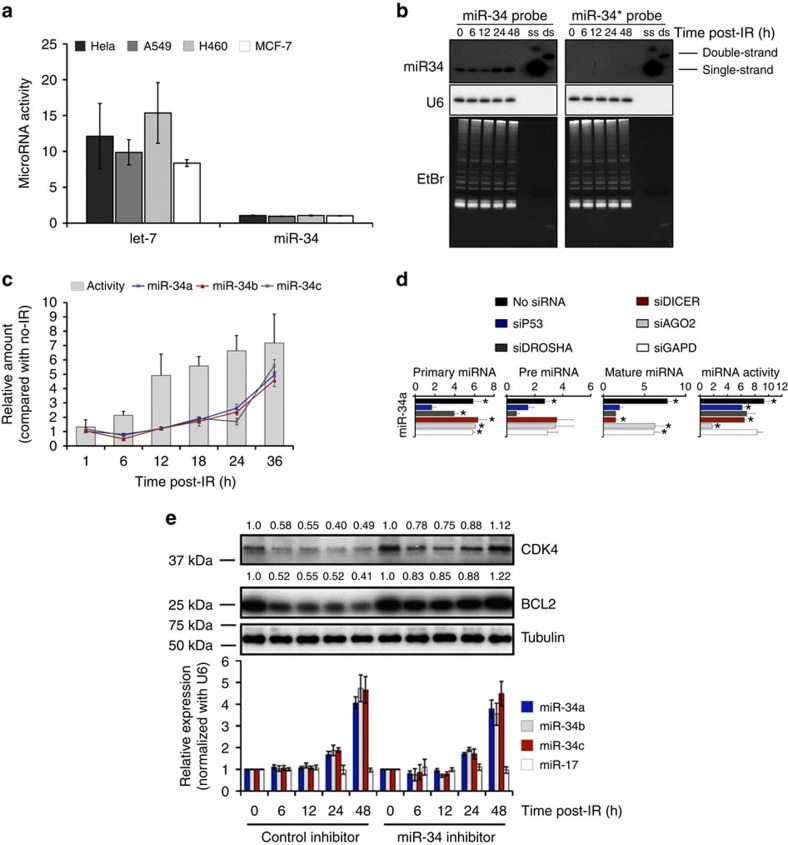
Figure 1. DNA damage activates a pool of existing, mature miR-34 that leads to strong gene repression.
(a) miR-34 and let-7 activity in cells 16 h after transfection with dual-luciferase psi-miR-34 (WT or MT) and psi-let-7 (WT or MT) reporters. Renilla was normalized to Firefly and miRNA activity was expressed as the fold-suppression of MT/WT. Graphed is the average±s.d. of three experiments in triplicate. (b) Northern blot of miR-34 and miR-34* from A549 cells exposed to 6 Gy of IR. U6 and ethidium bromide-stained gels were used as a loading control. (c) A549 cells expressing psi-miR-34 (WT or MT) reporters were lysed at the indicated time after exposure to 4 Gy. Lysates were assayed for dual-luciferase and mature miR-34a/b/c expression by RT–qPCR. Renilla was normalized to Firefly and was expressed as the fold-suppression of MT/WT. MiR-34 expression was normalized to U6. Data are expressed as the average fold change±s.d., relative to non-irradiated cells (lysed in tandem) of three independent experiments. (d) A549 cells expressing the psi-miR-34 (WT or MT) reporters were transfected with siRNA and lysed 36 h after exposure to 4 Gy. Lysates were analysed for luciferase, pri-miR-34a, pre-miR-34a and mature miR-34a expression by RT–qPCR. Renilla was normalized to Firefly. Pri-miR-34a and pre-miR-34 were normalized to β-actin mRNA, mature miR-34a was normalized to U6. Graphed is the fold change±s.d., relative to non-irradiated cells; n=4 independent experiments. *P<0.05, one-tailed Student's t-test. (e) A549 cells transfected with 2′-O-methyl inhibitors were exposed to 6 Gy of IR. Cells were lysed at the indicated time post IR. Lysate was split and analysed for protein expression by western blot (top) and miR-34 expression (bottom) by RT–qPCR. Bands were quantificated using ImageJ.

