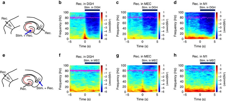
Figure 3. Selectively activating GABAergic interneurons suppress ictal seizure activities.
(a,e) The targeting sites for optical stimulation and electrical recording in kainite mice. (b–d,f–h) Mean spectrograms of LFPs recorded in the DGH (b,f), MEC (c,g) and M1 (d,h), respectively (n=10 mice). Data were normalized to the maximal log10(power) value across the whole 4–100 Hz frequency interval. Purple traces are raw LFP data from representative channels recorded in the DGH (b,f), MEC (c,g) and M1 (d,h), respectively. Light pulses (473 nm, 5 ms pulse duration at 130 Hz) were delivered into the DGH (a–e) and MEC (f–j) at time 0. The thick blue line denotes the initial 5 s of stimulation periods.

