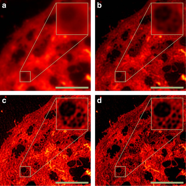
Figure 4. SR-SIM measurement of an LSEC membrane stain obtained on a GE Healthcare DeltaVision|OMX.
Wide-field image (a), Wiener-filtered wide-field (b), single-slice/2D SR-SIM reconstruction by fairSIM (c) and full 3D SR-SIM reconstruction by SoftWORX (manufacturer's software) (d) are shown for comparison. Both SR-SIM reconstructions allow to clearly identify the cell's fenestrations (tiny membrane pores), which is not possible in the wide-field images. The single-slice reconstructions by fairSIM can be performed with a much lower number of input images, as no z-stack has to be acquired. Scale bar, 5 μm, inset 1.6 μm, cells stained with CellMask Deep Red.

