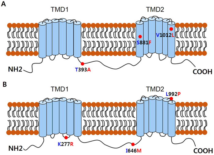
Figure 3. Schematic diagrams of the predicted structures of the MDR50 and MDR65 proteins, each encompassing twelve transmembrane segments in two TMDs.

(A) The MDR50 protein had the T393A, S881F, and V1012L amino acid replacement sites. (B) The MDR65 protein had the K277R, I646M, and L992P sites. Amino acid replacements that may result in structural changes are indicated by red dots. Blue letters represent 91-C; red letters represent 91-R.
