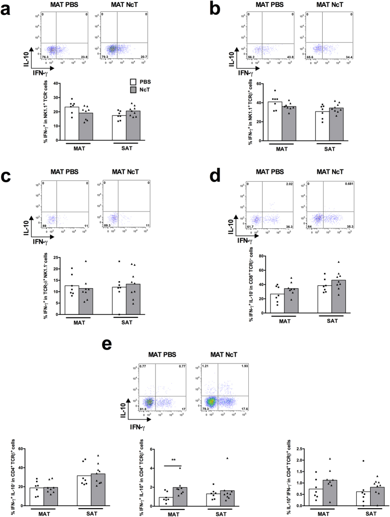
Figure 2. Impaired production of IFN-γ in the adipose tissue of infected IL-12/IL-23 p40−/− mice.
Frequencies of (a) IFN-γ+ NK1.1+ TCRβ−TCRγδ− cells on total NK1.1+ cells, (b) IFN-γ+ NK1.1+ TCRβ+ TCRγδ− cells on total NK1.1+ TCRβ+ cells, (c) IFN-γ+ TCRγδ+ NK1.1− cells on total TCRγδ+ cells, (d) IFN-γ+ IL-10−CD8+ TCRβ+ TCRγδ−NK1.1− cells on total CD8+ T cells and (e) IFN-γ+ IL-10−CD4+ TCRβ+ TCRγδ−NK1.1−, IFN-γ+ IL-10+ CD4+ TCRβ+ TCRγδ−NK1.1− and IL-10+ IFN-γ−CD4+ TCRβ+ TCRγδ−NK1.1− cells on total CD4+ T cells in the mesenteric and subcutaneous adipose tissue (MAT and SAT, respectively) from IL-12/IL-23 p40−/− mice sacrificed 24 h after intraperitoneal challenge with 1 × 10⁷ N. caninum tachyzoites (NcT) or PBS, as indicated. Each symbol represents an individual mouse. Bars represent means of 7–9 mice per group pooled from 2 independent experiments. (Mann-Whitney U, **P ≤ 0.01). Representative example of gating strategy used to define the respective cellular populations in the different depots of adipose tissue analysed. The example shown corresponds to MAT.

