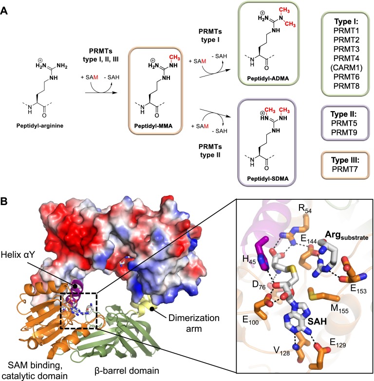
Figure 2.
Structure and mechanism of PRMTs. (A) Schematic representation of the PRMT catalyzed arginine methylation reactions including the different types of PRMTs mediating these enzymatic reactions. The classification of individual PRMT members is shown on the right side. (B) The crystal structure of dimeric PRMT1 bound to SAH and arginine (PDB code: 1OR8). The protomer on top is shown as surface representation colored according to its electrostatic potential (negative electrostatic potentials are highlighted in red, whereas positive electrostatic potentials are illustrated in blue). The inset on the right depicts a close up view of the PRMT1 active site residues implicated in substrate and cofactor binding as well as catalysis.

