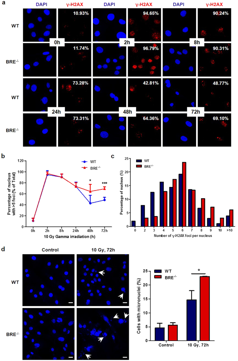
Figure 6. The DNA repair process is impaired in BRE−/− fibroblasts.
(a) Immunofluorescence staining of passage 4 WT and BRE−/− fibroblasts at indicated time points after 10 Gy of gamma irradiation using anti-γ-H2AX antibody (red), together with DAPI nuclear staining (blue). (b) Quantification of nuclei with more than five γ-H2AX foci in percentage at each time point for the WT and BRE−/− fibroblasts above. At least 150 cells were scored for the γ-H2AX foci in no fewer than five fields of duplicate plates. Note the significantly longer persistence of γ-H2AX foci in BRE−/− fibroblasts compared with WT fibroblasts. Data represent the mean ± SD. *denotes P-value < 0.05, ***denotes P-value < 0.001 versus WT. (c) Tabulation of the percentage of nuclei containing indicated numbers of γ-H2AX foci at 72 h after irradiation. (d) DAPI nuclear staining showing more cells with fragmented micronuclei among the BRE−/− compared with WT fibroblasts at 72 h after irradiation. Arrowheads indicate examples of micronuclei (left panel). Scale bars = 20 μm. Quantification of cells with micronuclei (right panel). At least 300 were counted in no fewer than five fields of three independent experiments. Data represent as the mean ± SD *denotes P-value < 0.05 versus WT.

