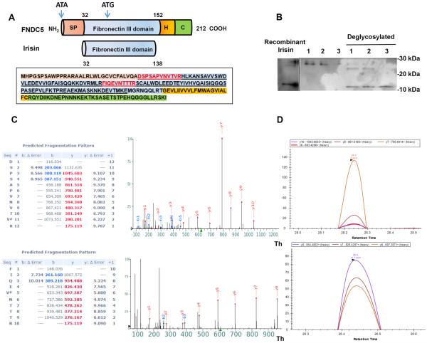
Figure 1. Analysis of Irisin Peptides by Mass Spectrometry.
(A) Schematic representation of the FNDC5 protein structure (top) and irisin (bottom). SP = signal peptide, H = hydrophobic domain, C = c-terminal domain. Human FNDC5 sequence with corresponding domains colored. Human irisin sequence is underlined as well as synthetic AQUA peptides used in this study (red).
(B) Immunoblotting of irisin plasma samples from three subjects undergoing aerobic interval training with or without deglycosylation enzyme (Protein Deglycosylation Mix (NEB)) and deglycosylated recombinant irisin.
(C) MS2 spectra acquired using a Q Exactive mass spectrometer for the two synthetic AQUA peptides and their b-, y-ion series m/z values. Mass accuracy values are given in PPMs and “#” denotes the heavy valine residue.
(D) PRM elution profile for the y-ions for the AQUA peptides using Skyline software. Retention times for each peptide are labeled on the x-axis and y-axis represents the relative intensity for each y-ion peak. See also Figure S1.

