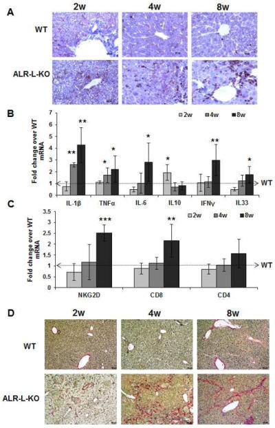
Figure 3. Hepatic inflammation and fibrosis in ALR-L-KO mice.

(A) CD45 staining indicates progressively increased inflammatory cells in the ALR-L-KO liver. (B) Hepatic mRNA expression of inflammatory cytokines (IL1β, TNFα, IL6, IFNγ and IL33) increased strongly at 8 weeks in ALR-L-KO relative to WT mice. Anti-inflammatory IL10 increased at 2 weeks and declines to the WT level at 4 and 8 weeks. (C) Hepatic mRNA expression of NKG2D and CD8, but not CD4, increased at 8 weeks. *P<.05, **P<.01 and ***P<.001 vs expression in WT liver. (D) Sirius Red staining shows mostly pericellular fibrosis at 2 weeks and periportal fibrosis at 4 weeks, whereas at 8 weeks there is bridging fibrosis.
