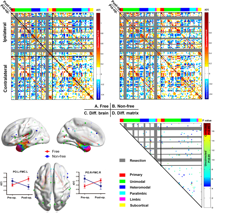
Figure 4. Interaction effect (treatment by outcome) for network edges.
(A,B) The pre- (lower diagonal) and postoperative (upper diagonal) correlation matrix for seizure-free (A) and non-seizure-free groups (B). (C) Two connections (edges) with significant interaction effects are shown in a 3D brain template (P < 0.05 corrected) by line segment in red. Line graphs show how these connections were altered by surgery in each group. (D) Matrix showing connections with significant interaction effect, both corrected (in red) and uncorrected (colored blue to yellow). Columns and rows (nodes) of the matrices are ordered and colored according to six distinct functional modules (Fig. 1, Table S1). The nodes damaged by surgery are shown in gray. PO.L = left parietal operculum cortex, FMC.L= left frontal medial cortex, PO.R = right parietal operculum cortex, SC.R = right subcallosal cortex, PO.L = left parietal operculum cortex.

