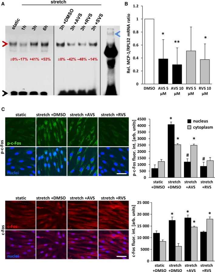
Figure 1.

Statins inhibit stretch‐induced activation of AP‐1 in HUVSMCs. A, Nuclear extracts from HUVSMCs, stretched for 1 to 6 hours and/or treated or pretreated with 10 μmol/L AVS, RVS, or SVS, were exposed to 32P‐labeled double‐stranded gel shift ODNs mimicking a consensus AP‐1 DNA binding site, separated by nondenaturing polyacrylamide gel electrophoresis and subjected to autoradiography (red arrowhead: AP‐1–ODN/protein complex; black arrowhead: unbound AP‐1–ODN; blue arrowhead: band of the AP‐1–ODN/protein complex supershifted by using an anti–c‐Jun antibody). The percentaged alterations in the band intensities are indicated with static or stretch set to 0%. B, mRNA expression ratios of MCP1 and RPL32 (housekeeping gene) in statin‐treated HUVSMCs were determined by real‐time polymerase chain reaction. Control conditions (DMSO) were set to 1 (*P<0.05/**P<0.01 vs DMSO, n=3). C, HUVSMCs were exposed to biomechanical stretch for 24 hours with or without statin pretreatment (1 hour at 10 μmol/L). Abundance of the AP‐1 subunit c‐Fos (red fluorescence) and activated (phosphorylated) c‐Fos (green fluorescence) in the nuclei (blue fluorescence, black bars) and cytoplasm (gray bars) was determined by quantitative immunofluorescence analyses (*P<0.05 vs static plus DMSO, # P<0.05 vs stretch plus DMSO; bars represent the mean±SD of values obtained from 5 microscopic fields of view of 1 of 2 experiments with comparable results; scale bars = 50 μm). arb. unit indicates arbitrary unit; AP‐1, activator protein 1; AVS, atorvastatin; DMSO, dimethyl sulfoxide; fluor. int., fluorescence intensity; h, hour; HUVSMC, human umbilical vein smooth muscle cell; ODN, oligodeoxynucleotide; p‐c‐Fos, phosphorylated c‐Fos; Rel., relative; RVS, rosuvastatin; SVS, simvastatin.
