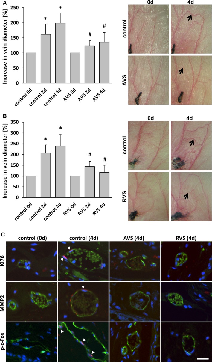
Figure 8.

Treatment with AVS or RVS blocks varicose‐like venous remodeling in the mouse auricle model. Daily imaging of the vasculature of the mouse auricle for 4 days after ligation of a central vein revealed increased tortuosity and the growth (A and B, arrows) of connected small collateral veins; this was significantly inhibited by systemic administration of AVS or RVS. A and B, *P<0.05 vs control day 0, # P<0.05 vs corresponding control day 2/4, n=5–7. Representative images show the venous network of mouse auricles (A and B) and cross‐sections of corresponding blood vessels (C) (confocal images; green fluorescence: CD31) showing Ki67‐positive nuclei (arrowheads), MMP2 accumulation (arrowhead), and p‐c‐Fos–positive nuclei (arrowheads) in remodeling veins (red fluorescence). Corresponding activity was not observed in veins from statin‐treated mice (scale bars = 20 μm). AVS indicates atorvastatin; d, days; p‐c‐Fos, phosphorylated c‐Fos; RVS, rosuvastatin.
