
Figure 3.
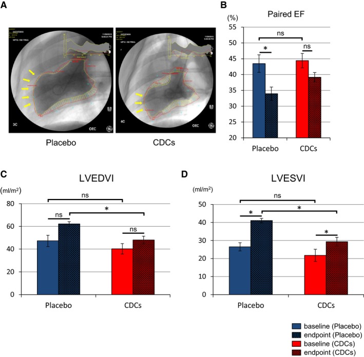
Allogeneic CDCs preserve cardiac function and attenuate adverse remodeling compared to placebo pigs, as assessed by left ventriculography. Representative cine pictures of left ventriculography in placebo and CDC‐treated pig at endpoint (2 months; A). Yellow arrow indicates akinetic anterior wall of left ventricle. Paired EF in each groups at baseline (≈15 minutes postreperfusion) and endpoint (B). Paired end‐diastolic volume index (C) and end‐systolic volume index (D) in placebo and CDC groups at baseline and endpoint. CDCs indicates cardiosphere‐derived cells; EF, ejection fraction; LVEDVI, left ventricular end‐diastolic volume index; LVESVI, left ventricular end‐systolic volume index. Values are means±SEM. *P<0.05 between 2 groups.