
Figure 4.
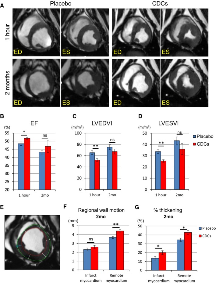
Allogeneic CDCs exhibit improved global function and remodeling 1 hour after cell treatment compared to placebo, as assessed by MRI. Matched cine short‐axis images (at end‐diastole [ED] and end‐systole [ES]) at 1 hour and 2 months post‐treatment for pigs treated with placebo and allogeneic CDCs (A). Comparison of ejection fraction (B), LVEDVI (C), and LVESVI (D) at 1 hour and 2 months in placebo‐ and CDC‐treated pigs. Endocardial (red) and epicardial (green) contours of the LV are shown. Cardiac slices were divided into 6 segments to calculate regional wall motion and thickening in infarct and remote myocardium at 2 months (E). Regional wall motion (F) and percent thickening (G) in infarct and remote myocardium in placebo and CDC groups at 2 months. CDCs indicates cardiosphere‐derived cells; EF, ejection fraction; LVEDVI, left ventricular end‐diastolic volume index; LVESVI, left ventricular end‐systolic volume index; MRI, magnetic resonance imaging. Values are means±SEM. *P<0.05 and **P<0.01 between 2 groups.