
Figure 3.
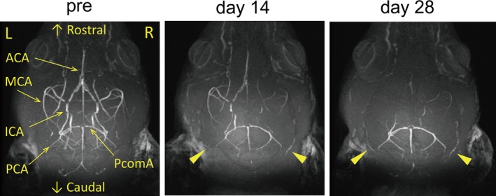
Gradual reductions of intracranial arterial flow signals after gradual common carotid artery stenosis (GCAS). Representative images of intracranial arterial flow signals obtained from 7‐T brain magnetic resonance angiography before GCAS (left), and at 14 days (middle) and 28 days (right) after GCAS. Arrowheads indicate gradual increment of collateral flow signals from posterior cerebral artery (PCA) to anterior circulation. ACA indicates anterior cerebral artery; ICA, internal carotid artery; MCA, middle cerebral artery; PcomA, posterior communicating artery.