
Figure 3.
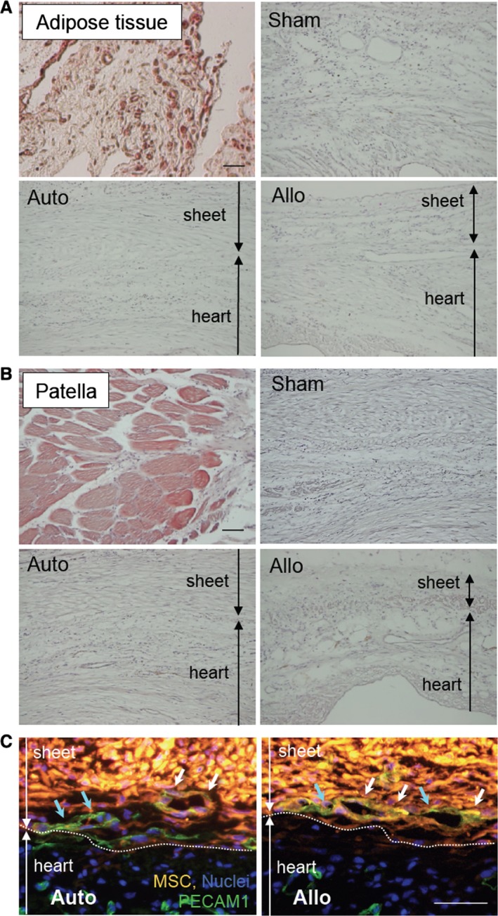
Donor MSC differentiation in vivo. A, Oil Red O staining detected the existence of adipocytes (dark red signal) in the peritoneal adipose tissue (positive control). No such Oil Red O–positive adipocytes were observed in the heart or in the MSC sheets at day 28 in any groups (5 hearts in each group). B, Alizarin red staining showed the calcium deposition in patella (positive control; red signal). No such signal was observed throughout the hearts or in the sheets at day 28. C, Immunohistostaining showed that there were DiI+ PECAM1+ donor‐derived endothelial cells (white arrows) as well as DiI− PECAM1+ host‐derived endothelial cells (blue arrows) in the sheets at day 3 similarly in the auto and allo groups. Green indicates PECAM1; orange indicates donor MSCs (CM‐DiI); blue indicates nuclei (DAPI). For (A through C), 5 hearts were studied in each group. Scale bars=100 μm (A and B) and 50 μm (C). Allo indicates allogeneic MSC sheet placement at 4 weeks after myocardial infarction; Auto, syngeneic MSC sheet placement at 4 weeks after myocardial infarction; MSC, mesenchymal stromal cell; Sham, sham treatment (open chest only) at 4 weeks after myocardial infarction.