
Figure 10.
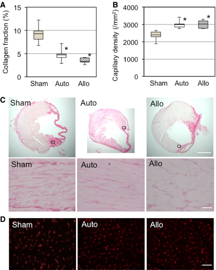
Allogeneic MSC sheets induced myocardial repair comparable to syngeneic MSC sheets. A, Extracellular collagen deposition in the peri‐infarct area, assessed by picrosirius red staining at day 28, was reduced in the auto and allo groups compared with the sham group. B, Isolectin B4 staining detected an increase in capillary density in the peri‐infarct area in the auto and allo groups compared with the sham group at day 28. C, Representative images for (A). Upper panels are whole heart images, whereas lower panels are higher magnification images of chosen areas (black squares) in the border area. Collagen deposition is shown in deep pink. Scale bar=300 μm (upper panels) and 50 μm (lower panels). D, Representative images for (B). Capillaries are stained in red. n=5 hearts per group. *P<0.05 vs sham. Scale bar=50 μm. MSC indicates mesenchymal stromal cell.