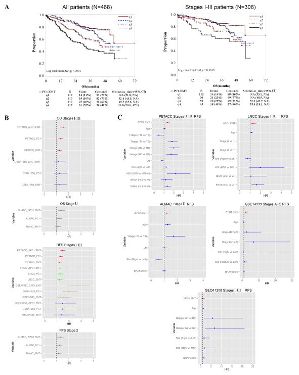
Figure 2. ΔPC1.EMT is associated with poor overall survival on the Moffitt468 CRC dataset and on independent CRC datasets.
(A) Kaplan Meier (KM) survival analysis of quartile scores on Moffitt468 shows that a higher ΔPC1.EMT predicted poorer overall survival (OS) for all 468 patients (left panel), and also when limited to the 306 stage I–III primary tumors (right panel). Also see Supplementary Figures S1–S3 for additional analyses on MSS, MSI, and metastatic tumors as well as for PC1 and EMT scores. (B) Forest plot summary of OS and RFS univariate analyses of EMT, PC1 and ΔPC1.EMT scores on PETACC3, LNCC, GSE14333, GEO41258 and ALMAC datasets. See Supplementary Table S4 for detailed information with other clinico-pathological and molecular variables. Note: signature scores were standardized by IQR. (C) Forest plot summary of RFS multivariable analyses of the ΔPC1.EMT score on PETACC3, LNCC, GSE14333, GEO41258 and ALMAC datasets. Note: the solid lines represent 95%CI and signature scores were standardized by IQR. See Supplementary Figure S4 for OS multivariable analyses. Also see Supplementary Tables S5–S9 for detailed information for PC1 and EMT scores as well as other clinico-pathological and molecular variables.

