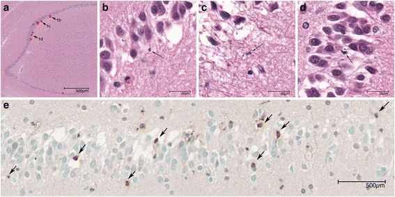
Fig. 1.

Apoptotic cells in hippocampus dentate gyrus. a Overview of hippocampus in HE stain. The apoptotic cells are marked with red dots. b–d Three apoptotic cells are indicated by arrows and magnified in b, c and d. Arrows again indicate the apoptotic cells. e. TUNEL analysis. Arrows indicate the positive cells
