Fig. 2.

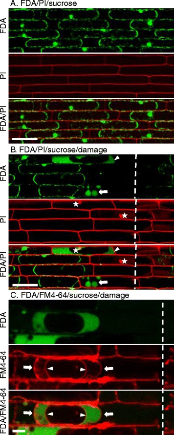
Novel fluorescein patterns in the cytoplasm of cells next to directly damaged cells. a Confocal image showing dual staining with FDA (green) and PI (red) followed by treatment with 0.5 M sucrose to induce plasmolysis in live cells. Bar = 50 μm. b Confocal image showing dual FDA/PI staining followed by treatment with 0.5 M sucrose to induce plasmolysis in live cells, then mechanically damaged with a razor. White dotted lines indicate where the sheath was damaged with the razor. White stars indicate PI stained nuclei. White arrows indicate membrane bound compartments containing fluorescein. Arrowheads indicate fluorescein evenly distributed in the cytoplasm but excluded from the vacuole. Bar = 50 μm. c Confocal image of rice cells sequentially treated with FM4-64 for two hours, FDA for 10 min and 0.5 M sucrose for 10 min, followed by mechanical damage with a razor. White dotted lines indicate where the cell was damaged with the razor. FM4-64 stained the plasma membrane (arrow) and the tonoplast (arrowhead), and fluorescein (green) was retained in the cytoplasm. Bar = 10 μm
