Fig. 3.

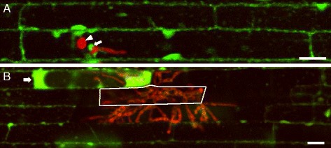
Host cell viability at early and late stages of rice blast invasion. a Single plane confocal image of rice sheath epidermal cells infected with M. oryzae transformant CKF1997 expressing cytoplasmic tdTomato (shown in red) at 28 hpi and stained with FDA (green). The appressorium (arrowhead) mediated penetration of the host cell and produced IH. Fluorescein is localized in the cytoplasm of both infected and non-infected cells and also associated with a BIC (arrow). b Maximum projection of three successive z-stack images covering 6 μm, showing rice sheath epidermal cells infected with M. oryzae transformant CKF1997 at 48 hpi and stained with FDA. IH (red) had spread into two cells away from the initially invaded cell indicated with solid white outline. Newly invaded- and non-invaded cells were stained with fluorescence, whereas fully invaded and some partially invaded cells lacked fluorescein. A novel fluorescein pattern (brighter fluorescence in the enlarged cytoplasm) was observed in a partially invaded cell (white arrow). Bars = 20 μm
