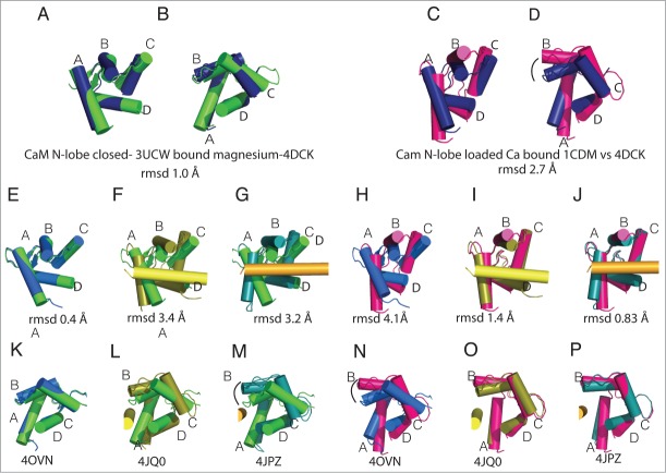
Figure 4.
CaM N-lobe conformations in Nav-CaM complexes. The structures of the CaM N-lobe in the different complexes are compared with the isolated CaM N-lobe with Mg2+ (green, 3UCW 40,41) or CaM-Ca (magenta, 1CDM 40). Helix αVI is shown in the structures in which there is an interaction between the CaM N-lobe and the helix. In the figure, CTNav1.5-CaM-FHF (navy blue, PDB ID 4DCK), CTNav1.5-CaM (marine, PDB ID 40VN), CTNav1.5-CaM-Ca-FHF (olive, PDB ID 4JQ0), CTNav1.2-CaM-Ca-FHF (teal, PDB ID 4JPZ), and CaM-Ca (magenta, PDB ID 1CDM) are shown. (A) Overlap of the CaM N-lobe as seen in CaM N-lobe with Mg2+ (PDB ID 3UCW) with CTNav1.5-CaM-FHF (navy, PDB ID 4DCK). (B) 90° rotation of A. (C) Overlap of the CaM N-lobe as seen in CaM-Ca (PDB ID 1CDM) with CTNav1.5-CaM.FHF (navy). (D) 90° rotation from C. (E, K) Alignment of the CTNav1.5-CaM N-lobe with CaM N-lobe-Mg (rmsd 0.4 Å). (H, N) Alignment of the CTNav1.5-CaM with CaM-Ca (rmsd 4.1 Å). (F, L) Alignment of the CTNav1.5-CaM-Ca-FHF N-lobe with CaM N-lobe-Mg (rmsd 3.4 Å). (I, O) Same as (F, L)with CaM-Ca (rmsd 1.4 Å). G, M. Alignment of the CTNav1.2-CaM-Ca-FHF N-lobe with CaM N-lobe-Mg (rmsd 3.2 Å). (J, P) Same as (G, M)with CaM-Ca (rmsd 1.45).

