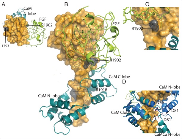
Figure 8.
Location of mutations of Nav1.2 observed in autism and febrile seizures. (A) Surface representation of CTNav1.2 (top view, orange) interacting with CaM (teal) and FHF (lime green). Mutation of residue 1793 of the EFL domain is shown in gray. (B) CTNav1.2 mutations in gray (1902, 1918). (C) Close-up of residue R1902 close to the interface of CTNav1.2 with FHF. (D) Close-up of the position of residues Asp79 and Asp81 of CaM in the CTNav1.2-CaM-Ca-FHF (antiparallel; teal) vs CTNav1.5-CaM (parallel; marine).

