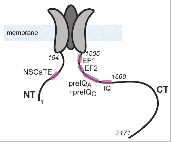
Figure 1.

Illustration of the structure of α1C subunit of CaV1.2. NT and CT and 3 out of the 4 transmembrane domains of α1C are shown schematically. Cytosolic loops I, II and III are not shown. In italics are numbers of selected a.a. residues in NT and CT, shown for orientation.
