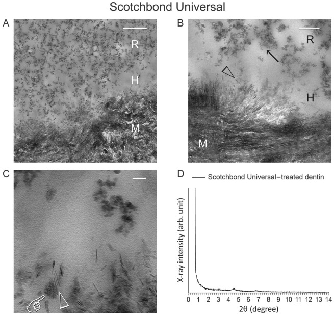
Figure 3.
Representative example of resin-dentin interface created by a commercial adhesive that contains 10-methacryloyloxydecyl dihydrogen phosphate (10-MDP) (Scotchbond Universal; pH 2.7) in coronal dentin, in which no nanolayering could be identified in any location or section. (A) Low-magnification transmission electron microscopy (TEM) image taken from an unstained, nondemineralized section. A 0.5-µm-thick hybrid layer (H) is present between the filled adhesive resin (R) and mineralized dentin (M). Bar = 500 nm. (B) Remnant apatite crystallites (open arrowhead) are present within the hybrid layer (H). Nano-sized filler clusters (arrow) can be found in adhesive resin (R). Bar = 200 nm. (C) High-magnification image showing formation of mineral salts (pointer) around remnant apatite crystallites (open arrowhead), with no evidence of nanolayering. Bar = 50 nm. (D) Thin-film glancing angle X-ray diffraction (XRD) spectrum from 2° to 14° 2θ of Scotchbond Universal–treated dentin. The scan was plotted using the same horizontal and vertical scales as Figure 2D. Characteristic peaks of 10-MDP-calcium salt can barely be discerned. M, mineralized dentin.

