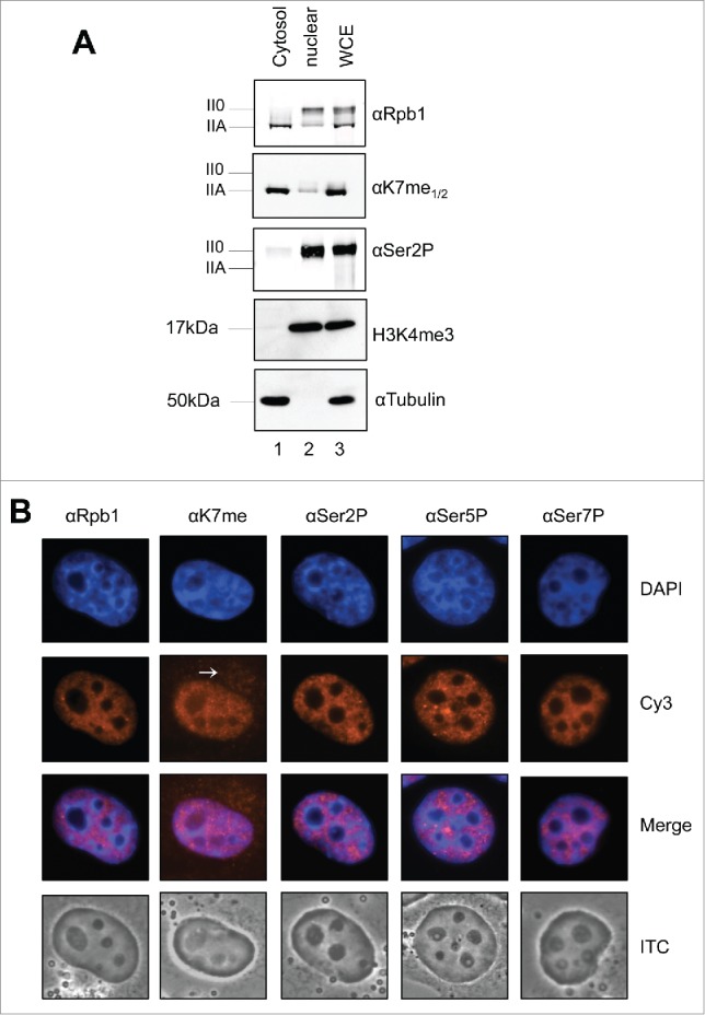
Figure 3.

Cytoplasmic - nuclear distribution of Kme1/2 methylated RNAPIIA. (A) Cytoplasmic and nuclear extracts of H1299 cells were prepared and analyzed in Western blots. The ratio of RNAPIIA to RNAPII0 in the cytoplasm and nucleus was visualized with mAbs Pol3.3 and Ser2P. H3K4me3 served as nuclear markers and tubulin as cytoplasmic marker. (B) Immunofluorescence staining of formaldehyde fixed H1299 cells. Ser2P, Ser5P, and Ser7P modified CTD was observed exclusively in the nucleus, while significant CTD Kme1/2 staining was observed also for the cytoplasm. Merge: overlay of DAPI and Cy3; PhC: phase contrast.
