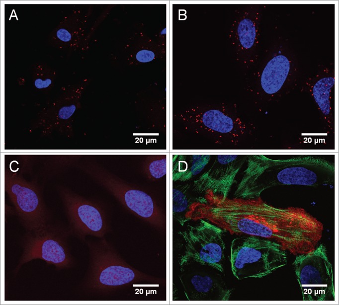
Figure 2.

In situ PLA confirms the interaction between Rab7b and myosin II in DCs and U2OS cells. (A) Monocyte-derived dendritic cells were fixed and stained with antibodies against Rab7b and myosin II, combined with secondary PLA probes (Duolink, Sigma). The interaction events are visible as red dots. The nuclei are stained in blue (Hoechst). Scale bar 20 μm. (B) U2OS cells transiently transfected with HA-tagged Rab7b were fixed and stained with antibodies against Rab7b and myosin II, and further treated with secondary PLA probes (Duolink, Sigma). The interaction events are visible as red dots. Scale bar 20 μm. (C) U2OS cells transiently transfected with HA-tagged Rab7b were fixed and stained with antibodies against Rab7b and the early endosomal marker EEA1 as a negative control. No interaction events are visible after in situ PLA. Scale bar 20 μm. (D) U2OS cells transiently transfected with HA-tagged Rab7b were fixed and immunostained with primary antibodies against Rab7b and myosin II, followed by Alexa-555 (red) and Alexa-488 (green) conjugated secondary antibodies, respectively. Scale bar 20 μm.
