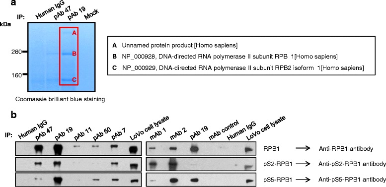
Fig. 2.

Identification of RPB1 as the antigen recognized by pAb 47, pAb 19, and mAbs. a LoVo cell lysates were immunoprecipitated with pAb 47 and pAb 19, and the immunoprecipitates were subjected to 4–12 % Bis-Tris gel electrophoresis. Gels were stained with Coomassie Brilliant Blue, the bands were excised, and their identities were determined using mass spectrometry. (b) LoVo cell lysates were immunoprecipitated with pAb or mAb (scFv-human Fc fusion protein). The resulting precipitates were subjected to immunoblot analysis with antibodies specific for RPB1 or phosphorylated RPB1. Normal human pooled IgG fractions and LoVo cell lysates were used as controls
